Death Under Glass: Vivid Images With A Dark Backstory
Last Updated 13 March 2015


Londonist Rating: ★★★★☆
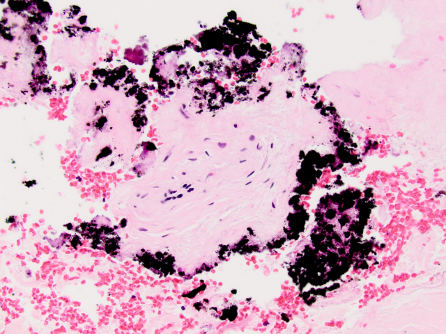
Forensic pathology is often portrayed as being cut and dry; all cold steel and clinical white lab coats. But it also has an intrinsic beauty and aesthetic — as portrayed in the photomicrographs of Death Under Glass — the current exhibition at Bart's Pathology Museum.
Dr. Marianne Hamel and forensic photographer Nikki Johnson have produced a unique vision of the post mortem, as viewed through the microscope. Their images focus on the cellular structures and stains used to identify specific anatomical or pathological features. And we reckon these 'artworks' rival any of those created by abstract painters.
The colours are vivid and bright, and it's fascinating to discover the causes behind them (fat embolisms, for example, are revealed through the bold red colouring of Oil Red O stain). Ironically, many of the stories behind the colours are dark ones; the lurid rainbow hues of fan-shaped oxalate crystals actually indicate fatal poisoning by antifreeze.
Death Under Glass is a deft fusion of art and science, and Bart's Pathology Museum, with its shelves of anatomical specimens, provides a fitting backdrop.
Dr. Hamel will also present a talk on her work on 15 April — it's an extra date, as an earlier talk has already sold out. .
Death Under Glass continues at Bart's Pathology Museum until April — admission times are limited. Check the website for further details. All images courtesy of Nikki Johnson and Dr. Marianne Hamel.